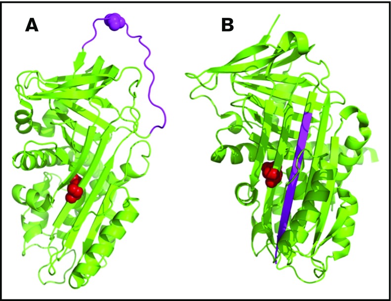
Figure 4.
Representative structure of the protein Z–dependent protease inhibitor (SERPINA10) showing the 3-dimensional orientation of residue Q384 (in red) and the reactive center loop (RCL, in pink). The protein model was created using PyMOL (PDB codes: 3H5C for native and 4AFX for RCL-inserted forms). (A) The native protein. (B) Binding of a target protease to the RCL results in cleavage and insertion of the RCL into the central β-sheet.

