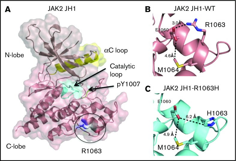
Figure 7.
Projected changes in JAK2 protein due to R1063H. Models were created by homology modeling on the SWISS-MODEL server and visualized using PyMOL (PDB 2B7A). (A) The JAK2-JH1 domain, with the catalytic loop highlighted in cyan, the αC loop in yellow, and phosphotyrosines within the activation loop in green. (B) In the wild-type protein, R1063 forms a salt bridge with E1060, which is exposed on the surface of the JH1 domain. (C) In the R1063H mutant protein, substitution of the charged arginine to a polar histidine results in the loss of the native salt bridge with El 060.

