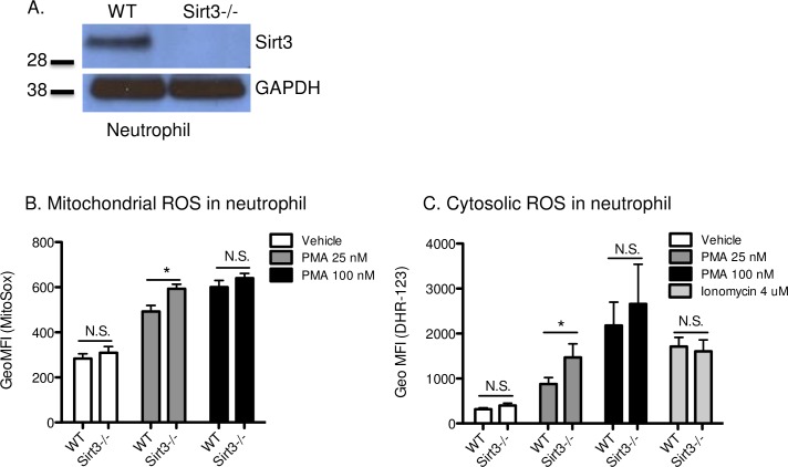
Fig 1. Deficiency of Sirt3 augmented production of ROS in neutrophils.
(A) Western blot analysis using bone marrow neutrophil lysates from Sirt3-/- mice or WT mice. Photograph of a representative blot is shown. (B) and (C) represent flow cytometric determination of mitochondrial and cytosolic ROS in neutrophils. Diluted anti-coagulated whole blood from Sirt3-/- mice and WT mice was incubated with (B), (C) PMA (25, 100 nM), (C) ionomycin (4 μM) or vehicle for 30 min at 37°C in the presence of (B) MitoSox or (C) dihydrorhodamine-123. MitoSox or Rhodamine-positive neutrophils were quantified by flow cytometry for each condition. n = 5–7. *P<0.05 vs WT (Student’s t tests).

