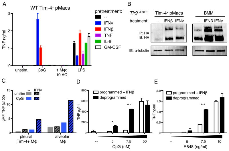
Figure 6. Inflammatory cues induce TLR9 expression in AC-engulfing macrophages.
(A) TNF production by Tim-4+ pMacs treated overnight with the indicated cytokines before stimulation with TLR ligands or ACs. Data are representative of at least three independent experiments. (B) Tim-4+ pMacs and BMMs from Tlr9HA:GFP mice were cultured overnight ± IFNγ or IFNβ, and TLR9 levels in lysates were measured by anti-HA immunoprecipitation and immunoblot. An anti-tubulin immunoblot was performed on lysates. Wells of Tim-4+ pMacs correspond to lysate of 1.6e6 cell equivalents, and wells of BMMs correspond to lysate of 0.4e6 cell equivalents. Data are representative of at least three independent experiments. (C) TNF production by lung and pleural cells harvested from WT mice and incubated overnight ± 100ng/ml IFNγ before stimulation with CpG ODN. Data are representative of two independent experiments. (D–E) TNF production by programmed Tim-4+ pMacs incubated overnight with IFNβ or by untreated deprogrammed Tim-4+ pMacs stimulated with increasing doses of CpG (D) or R848 (E). Data are representative of three independent experiments. See also Figure S6.

