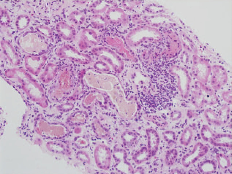
Figure 2.

Kidney section showing tubular atrophy, various casts (erythrocyte, leukocyte, and epithelial cell casts) in the distal tubules, and inflammatory cell infiltration in the interstitium (periodic acid–Schiff stain; original magnification 200×).
