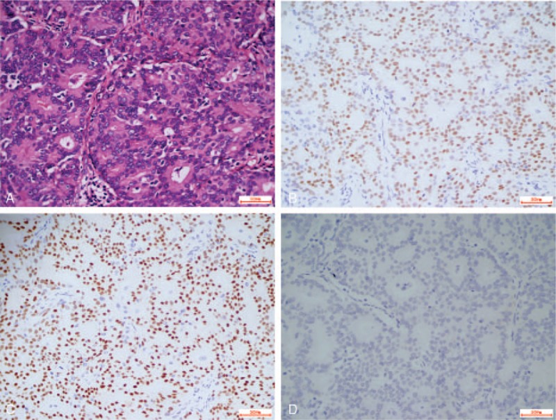
Figure 3.

(A) Histological appearance of invasive ductal carcinoma (hematoxylin and eosin stain, magnification ×400). Immunohistochemistry analysis showed that the tumor cells were positive for (B) estrogen receptor and (C) progesterone receptor, and (D) tumor cells were negative for HER2 (all, magnification × 400). HER2 = human epidermalgrowth factor receptor 2.
