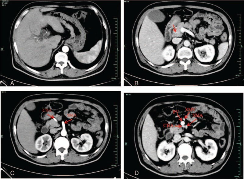
Figure 1.

Contrast-enhanced computed tomography. (A) Dilation of intrahepatic and extrahepatic bile duct. (B) The distal common bile duct was slightly enhanced in portal venous phase (arrow). (C) In arterial phase: the common hepatic artery originated from the superior mesenteric artery and cross between the pancreas head and the uncinate process. (D) In portal phase, CHA running posterior to the portal vein. CHA = common hepatic artery, PV = portal vein, SMA = superior mesenteric artery, SMV = superior mesenteric vein.
