Figure 1.

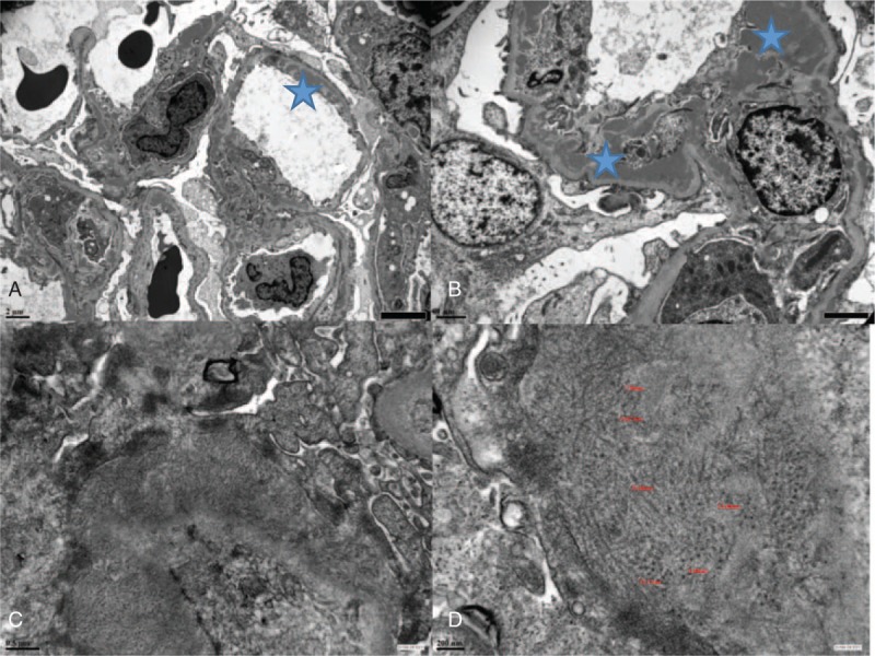
Electron microscopy. (A) Full House nephropathy, magnification ×5000: subendothelial electron-dense deposits in patient P4; (B) Full House nephropathy, magnification ×12,000: mesangial and subendothelial electron-dense deposits in patient P5; (C) fibrillary glomerulonephritis, magnification × 30,000: fibrils extending through the lamina densa of the glomerular basement membrane; (D) fibrillary glomerulonephritis, magnification × 60,000: deposits organized into unbranched, randomly oriented extracellular fibrils of 7–18 nm in diameter.
