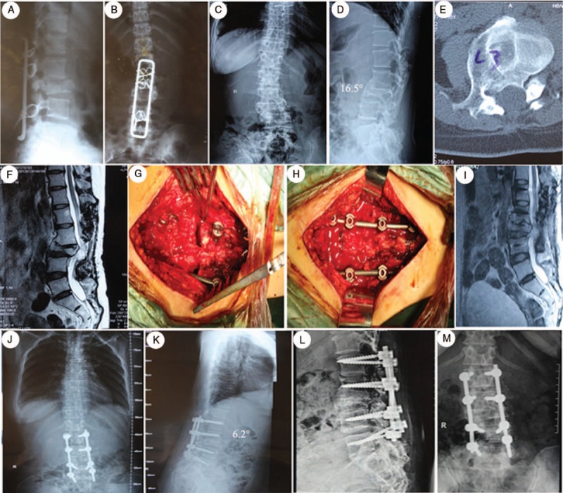
Figure 1.

A 35-year-old female who presented with an old L3 fracture and severe back pain. (A, B) She was treated with a posterior fusion and fixation from L1-L5, 14 years ago. (C, D) The preoperative x-rays showed the kyphotic angle was 16.5°, the lumbar lordotic angle was 9.2°. (E, F) Preoperative CT reconstruction and MRI scan showed bony canal stenosis and spinal cord compression. (G–H) Intraoperative pictures showed posterior osteotomy and fixation. (I) Postoperative MRI scan indicated complete decompression of the spinal cord. (J, K) She was treated with a posterior osteotomy and fusion from L1-L5 with L3 pedicle subtraction osteotomy. (L, M) A anteroposterior (AP) and lateral x-ray taken half a year after posterior fusion showed restoration of sagittal alignment without hardware loosening or loss of correction. CT = computed tomography, MRI = magnetic resonance imaging.
