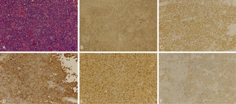
Figure 2.

Light microscopy and immunohistochemistry. (A) HE staining: the tumor cells were numerous small round cells with hyperchromatic nuclei (original magnification ×400). Immunohistochemistry showing (B) FLI-I (++), (C) INI-1 (++), (D) CD99 (+++), (E) S-100 (++), (F) Syn (++) (original magnification ×400). HE = hemotoxylin and eosin.
