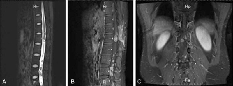
Figure 4.

A MR imaging before the second operation (9 months after the initial operation). MR imaging showing recurrence at T12-L1 levels and multiple seeding metastases at L3-L5 levels. (A) Sagittal T2-weighted MR image; (B) sagittal T1-weighted MR image; (C) coronal T1-weighted MR image. MR = magnetic resonance.
