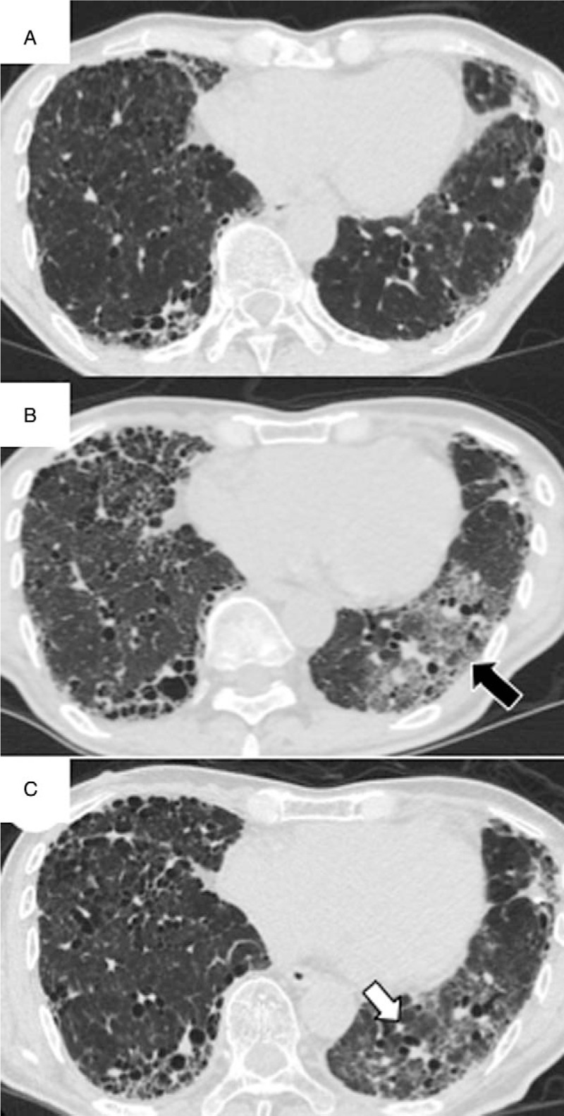
Figure 1.

Chest computed tomography (CT) findings for a 71-year-old man with idiopathic pulmonary fibrosis who presented with accelerated disease progression after nintedanib discontinuation. Chest CT findings at 8 months before admission (A), at the time of admission (B), and after treatment with methylprednisolone pulse therapy (1 g/d for 3 days) and prednisolone (C). Note the new ground glass opacities (GGOs) in the left lower lobe on images obtained at the time of admission (B, black arrow). After treatment, the GGOs were alleviated; however, the affected lobe exhibited volume loss with traction bronchiectasis (C, white arrow).
