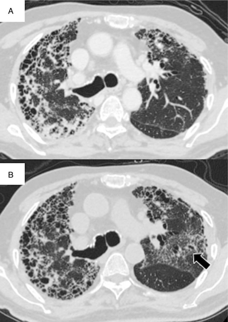
Figure 2.

Chest computed tomography (CT) findings for an 86-year-old man with idiopathic pulmonary fibrosis who presented with accelerated disease progression after nintedanib discontinuation. Chest CT findings at the time of nintedanib discontinuation (A) and at 3 weeks after discontinuation (B). Note the new ground glass opacities with traction bronchiectasis in the left upper lobe after discontinuation of nintedanib (B, arrow).
