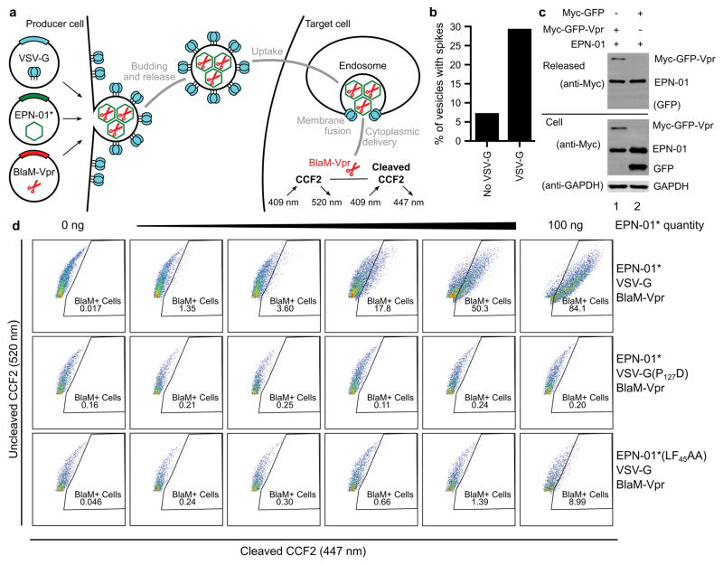
Extended Data Figure 7. EPNs can package biological cargoes and deliver them to the cytoplasm of target HeLa cells.
a, Schematic illustration showing the production, assembly, and release of EPNs incorporating BlaM-Vpr and VSV-G proteins (left), and detection of uptake and target cell membrane fusion using a BlaM colorimetric activity assay (right). b, Co-expression of VSV-G increases the number of vesicles that contain spikes, as evaluated by scoring >140 vesicles as either “containing” or “not containing” surface spikes (images like the one shown in the inset of Fig. 3b were scored independently by two different people, one blinded, and their counts were averaged). c, Western blots showing cellular expression and release of EPN-01 and Myc-tagged GFP constructs with or without fused Vpr (see Supplementary Table 3 for sequence information). Panel 1 shows released protein, panel 2 and 3 expression of Myc-tagged proteins and GAPDH in whole cell lysates. Lane 1 shows co-expression of EPN-01 with Myc-GFP-Vpr, lane 2 shows co-expression of EPN-01 with Myc-GFP. d, Flow cytometric analyses of HeLa cells loaded with the fluorescent CCF2 β-lactamase substrate and incubated with increasing quantities of wild-type EPN-01*/VSV-G/BlaM-Vpr (row 1), EPN-01*/VSV-G(P127D) mutant/BlaM-Vpr (row 2), and EPN-01*(LF45AA) mutant/VSV-G/BlaM-Vpr (row 3).

