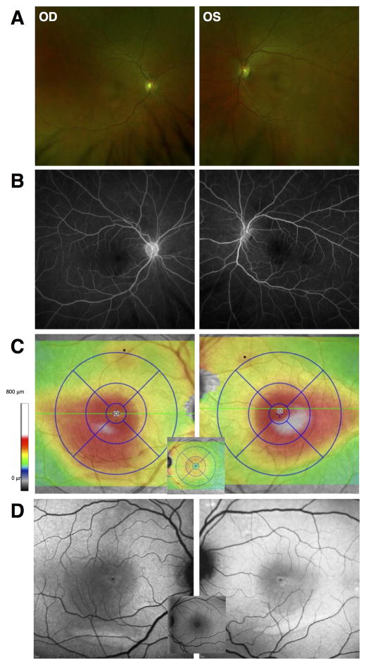
Figure 1.
A. Wide angle fundus photography showed a shallow, diffuse elevation of the macula and multifocal yellow-white depigmented lesions in both eyes. B. Mid-phase fluorescein angiography images did not show staining, pooling, or leakage in either eye. C. SD-OCT total retinal thickness topography maps showed increased overall retinal thickening extending from the foveal center to inferotemporal central retina in both eyes. D. Short-wavelength fundus autofluorescence (FAF) showed a diffuse increase in FAF that lightened the normally dark central region caused by macular pigment. Normal appearance is shown for reference as central insets in C and D.

