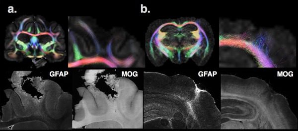
Figure 3.

Two examples of caveats are shown for the biophysical representation of diffusion MRI information by tractography in the ferret (a) and mouse (b) brain using the same approach and parameters. In the ferret brain near the site of a penetrating injury, FA is low and few tracts can be found in the body of the white matter compared with the contralateral side. However, investigation of this region using IHC of the same brain reveals the presence of myelinated axons (indicated by MOG IHC—myelin oligodendrocyte glycoprotein) and upregulated staining of astrocytes (by GFAP IHC—glial fibrillary acidic protein). The interpretation of the tractography in this case could indicate a loss of white matter fibers, when in fact the underlying pathology appears to be more related to gliosis. In the mouse brain (b), a region of increased FA and aberrant “tracts” can be found in the cortex near the injury site; however, inspection by IHC reveals a disruption of MOG staining and upregulation and organized GFAP staining in this tissue region of the same animal. The interpretation of tractography in this case could suggest cortical plasticity, when in fact the underlying alteration is more related to glial changes. This is similar to a finding reported by Budde et al. (2011). Taken together, this figure emphasizes the need for careful interpretation of dMRI findings, especially for biophysical models such as tractography, which directly report neurobiological metrics based model assumptions
