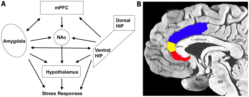
Figure 1.
Cortico-limbic circuitry implicated in mood regulation and depression. (A) Simplified schematic diagram of the cortico-limbic circuitry and the many interactions across the various brain regions. Not all known connections are depicted. Likewise, not all outputs of each region are depicted. mPFC, medial prefrontal cortex; HIP, hippocampus; NAc, nucleus accumbens. (B) Midline sagittal view of the human brain illustrating the location of major PFC regions, with the anterior cingulate cortex highlighted: blue, MCC24, mid-cingulate cortex; yellow, pACC24, pre-genual anterior cingulate cortex; red, SCC24/25, subcallosal cingulate cortex. Other brain regions noted include: dMF9, dorsomedial frontal cortex; vMF10, ventromedial frontal; OF11, orbitofrontal; A-Hc, amygdala-hippocampus in the temporal lobe; BS, brainstem; PCC23, posterior cingulate cortex; c. callosum, corpus callosum.

