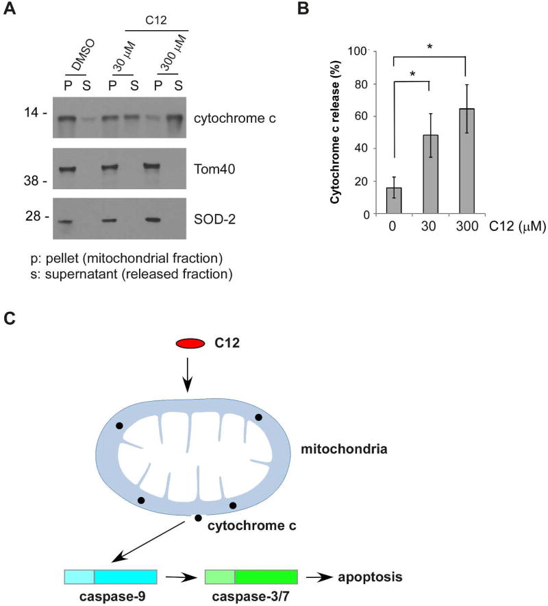
Figure 8. C12 directly induces mitochondrial outer membrane permeabilzation in vitro.
(A) Mitochondria were isolated from wild-type MEFs and incubated with C12 for 1 hour. Each sample contained 1% DMSO. Release of cytochrome c from mitochondria was determined by western blot. The molecular weight markers are labeled on the left (kD). (B) The intensities of cytochrome c in the mitochondrial (P) and the cytosolic (S) fractions shown in (A) were quantified using ImageJ. Cytochrome c release is represented as a percentage of the sum of the protein intensity in mitochondrial and released fractions. Mean ± standard deviation for four independent experiments are shown. Asterisks indicate P < 0.05 (*) by Student's unpaired t test. (C) C12 permeabilizes mitochondria, subsequently activating apoptotic signaling pathway.

