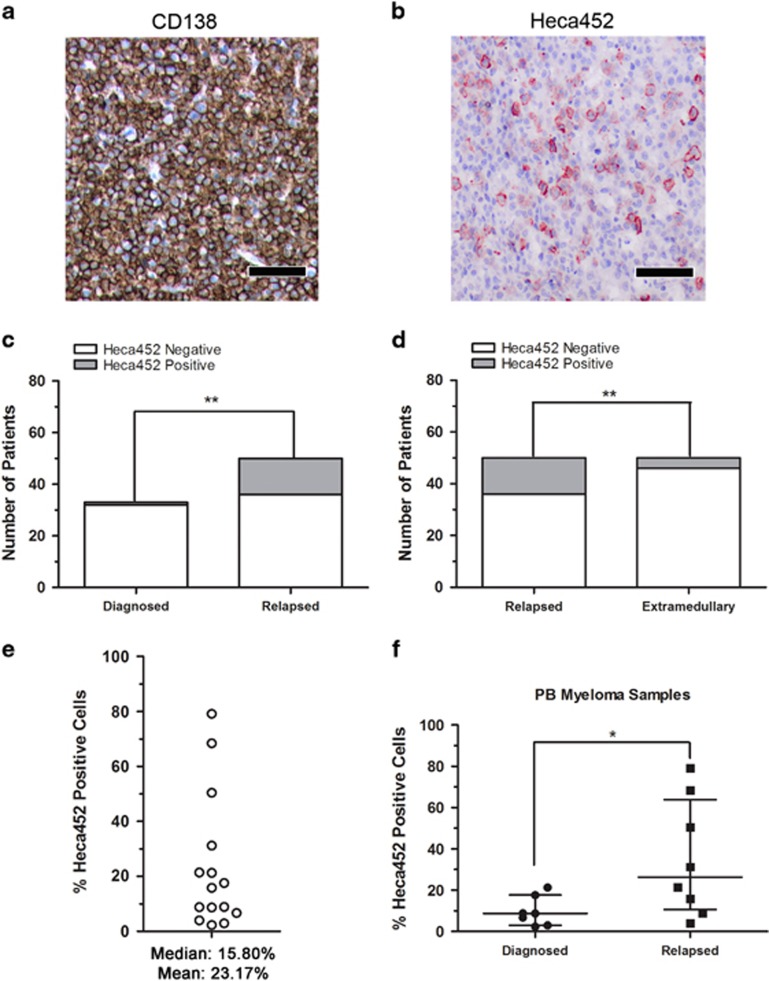
Figure 5.
Heca452-positive cells are present within the CD38/CD138 compartment of primary myeloma cells. Representative bone marrow sections of RRMM demonstrating a high-grade infiltration by neoplastic plasma CD138-positive cells (a) and Heca452 expression in a subpopulation of tumor cells (b). Bar represents 100 μm. Heca452-positive BM biopsies in newly diagnosed vs relapsed (c) and relapsed vs extramedullary (d) patients. The χ2-test was used to determine statistical significance in the Heca452-positive cells between groups of patients. ** and * represent P-values <0.01 and 0.05, respectively. (e) Quantification of the Heca452-positive cells by FACS. Cells were first selected based on their morphology (FSC-A/SSC-A) and viability status (7AAD negativity). Cells positive for CD2, CD14 and CD235a were excluded and CD38/CD138 double-positive cells were selected to screen for the presence of the Heca452-positive cells. Each empty circle represents percentage of Heca452-positive cells within the CD38/CD138 double-positive fraction for each PB samples from myeloma patients (N=15). (f) Comparison between the percentages of the Heca452-positive cells within the CD38/CD138 double-positive fraction from the PB of diagnoses vs relapse patients. Lines represent the median with interquartile range. Statistical significance was determined using the Mann–Whitney U-test. * represent P-values <0.05. FACS, fluorescence-activated cell sorter.

