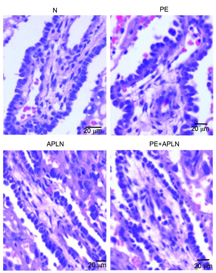
Figure 3.

Histology of placental tissue from rats (hematoxylin and eosin staining; scale bar, 20 µm). Compared with the placentas of N rats, those of PE rats had villous edema, irregularly arranged cells of various sizes and hyperchromatic nuclei, whereas PE+APLN rats had resolved edema of the placental villi and cells were regularly arranged with less hyperchromatic areas. N, control; APLN, apelin; PE, preeclampsia.
