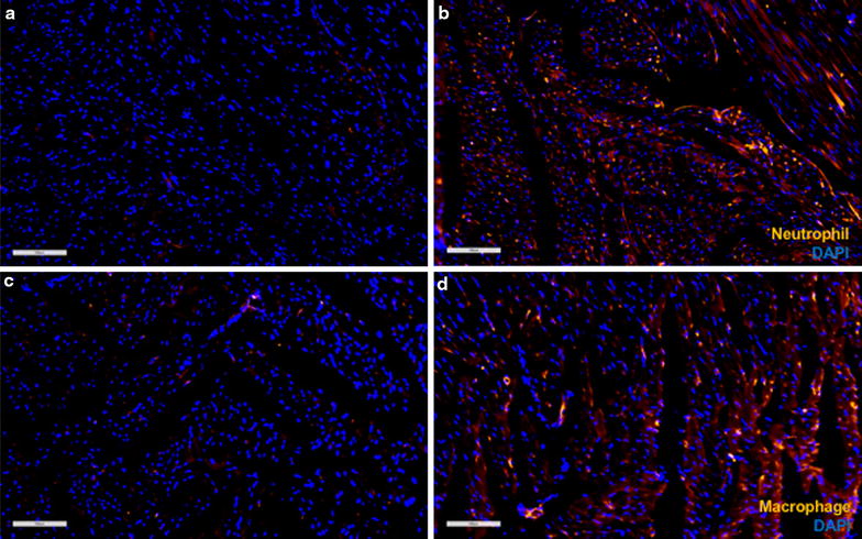
Fig. 8.

fIHC of macrophage and granulocyte infiltration into pFUS-targeted myocardium. a, b HIS48 staining showed more granulocytes in pFUS-targeted region compared to untreated regions. c, d CD68 staining showed more macrophages in pFUS-targeted region compared to untreated regions. Scale bar = 100 µm
