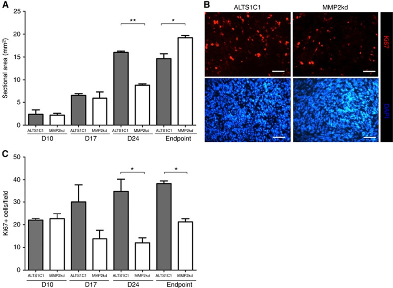
Figure 3.
MMP2 knockdown reduced the proliferation rate in vivo. (A) The change of tumour size was followed at the indicated day after tumour inoculation. The mean sectional areas of tumours were measured at the maximum cross section with H&E stain. (B) Representative images of Ki67 and DAPI staining in ALTS1C1 and MMP2kd tumours. Scale bar: 50 μm. (C) Quantification of Ki67+ cells was performed by counting the average of × 400 magnification field for sections in each tissue. *P<0.05, **P<0.01.

