Figure 5.
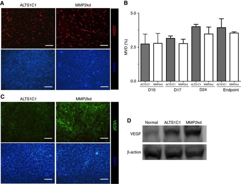
Suppression of MMP2 expression in tumour cells did not affect MVD, but increased VEGF expression. (A) Representative images of vessel density in tumours. Scale bar: 200 μm. (B) Quantification of MVD at the indicated day after tumour implantation. (C) Higher level of VEGF in MMP2kd tumours was detected by IHC staining. Scale bar: 200 μm. (D) The level of VEGF in normal brain, ALTS1C1, and MMP2kd tumour lysates was measured by western blotting.

