
Fig. 6.
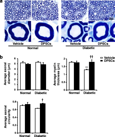
a Representative semi-thin cross-sections of the sural nerves of rats. Toluidine blue stained. b The morphometry of the myelinated nerve fibers. There were no significant differences in the diameter and circularity between the normal and diabetic rats. Myelin thickness was less in the diabetic rats compared with the normal rats. The transplantation of dental pulp stem cells (DPSCs) significantly increased the myelin thickness and circularity. The results are means ± SEM. **P < 0.01, vs. vehicle-injected side of normal rats; † P < 0.05, †† P < 0.01, vs. vehicle-injected side of diabetic rats; n = 5