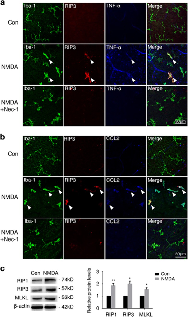
Figure 5.
Involvement of microglia necroptosis in the inflammatory process after acute retinal injury. For Nec-1 treatment in the NMDA-induced retinal injury mice model, NMDA and Nec-1 resolved in 1 μl PBS were injected into the vitreous of 8-week-old mice. Retinae were extracted 24 h later. (a and b) Immunostaining on retinal flat-mounts showed increased levels of RIP3, as well as inflammatory factors (TNF-α, CCL2) on Iba-1+ microglia after NMDA administration, whereas Nec-1 could decrease their expression, indicating the role of microglia necroptosis in the inflammation response. n=6 retinae per group. Scale bar: 50 μm. (c) Elevated protein levels of RIP1, RIP3, and their downstream MLKL from western blotting results confirmed the presence of necroptosis after NMDA damage. n=4 retinae per group. *P<0.05; **P<0.01

