Figure 7.
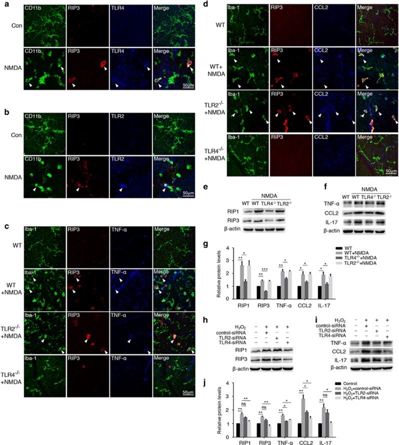
TLR4 activation rather than TLR2 was required in microglia necroptosis and inflammation. NMDA injury model was established on TLR2−/− and TLR4−/− mice. (a and b) Immunostaining on retinal flat-mounts showed distinct TLR2 and TLR4 activation in CD11b+ microglia from WT-NMDA mice (white arrows). (c and d) TLR4 knockout mice showed little RIP3 expression and Iba-1+ microglia co-labeled with either TNF-α or CCL2 was almost absent compared with WT-NMDA mice (white arrows), whereas in TLR2-deficient mice, Iba-1+ microglia displayed almost similar expression levels of RIP3, TNF-α and CCL2. n=6 retinae and eight images per retina. (e) Western blot revealed that RIP1 and RIP3 expression levels were reduced in TLR4−/− but not TLR2−/− mice after NMDA damage. (f) TLR4 knockout mice exhibited significantly impaired production of inflammatory cytokines (TNF-α, CCL2 and IL-17), whereas TLR2 deficiency showed nearly equal expression to WT mice. (g) Statistical analysis of western blotting are shown in e and f. n=4 retinae per group. (h) In vitro, BV2 cells transfected with either TLR2-siRNA, TLR4-siRNA, or control-siRNA were then stimulated with H2O2. Western blot showed decreased RIP1 and RIP3 expression in TLR4 rather than TLR2 knockdown cells. (i) Levels of inflammatory factors including IL-17, TNF-α and CCL2 were also significantly downregulated in TLR4-siRNA group compared with TLR2-siRNA and control-siRNA. (j) Statistical analysis of western blotting shown in h and i. n=4 retinae per group. *P<0.05; **P<0.01; ***P < 0.001; ns: no significance

