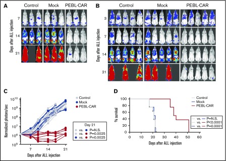
Figure 6.
PEBL-transduced T cells expressing an anti-CD7–41BB-CD3ζ CAR exert antitumor activity in xenografts. NOD-SCID-IL2RGnull mice were infused IV with 1 × 10⁶ CCRF-CEM cells labeled with luciferase. A total of 2 × 10⁷ PEBL-CAR T cells were administered IV on day 7 (A) or on day 3 and day 7 (B) after leukemic cell infusion to 3 and 5 mice, respectively. The remaining mice received either mock-transduced T cells or RPMI 1640 instead of cells (Control). All mice received 20 000 IU IL-2 once every 2 days IP. Shown is in vivo imaging of leukemia cell growth after d-luciferin IP injection. Ventral images of mice on day 3 in panel B are shown with enhanced sensitivity to demonstrate CCRF-CEM engraftment in all mice. The complete set of luminescence images is shown in supplemental Figure 7. (C) Leukemia cell growth in mice shown in panels A and B is expressed as photons per second. Each symbol corresponds to bioluminescence measurements in each mouse, normalized to the average of ventral plus dorsal signals in all mice before CAR T-cell infusion. (D) Kaplan-Meier curves show overall survival of mice in the different groups (8 in each group). Mice were euthanized when the total bioluminescence signal reached 1 × 10¹⁰ photons per second. The P values were calculated by log-rank test. N.S., not significant.

