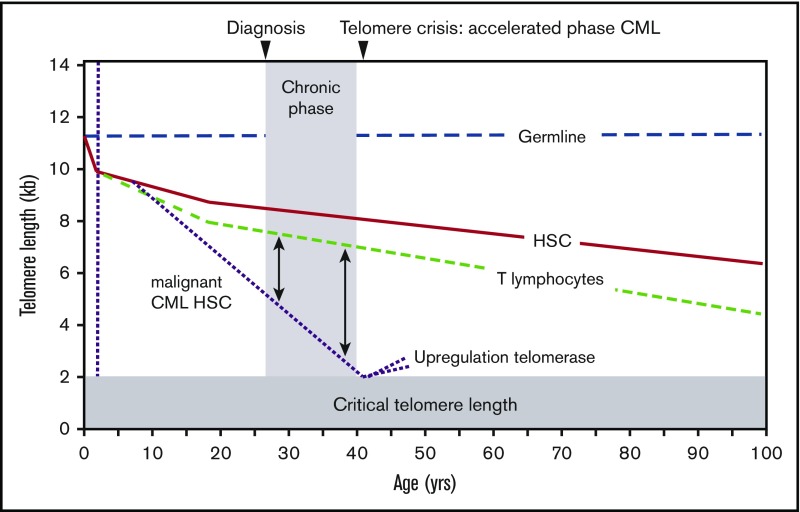
Figure 5.
Telomere length in CML. The difference in telomere length between CML blast cells relative to T lymphocytes in the same patient correlates with disease progression. See also Figure 3. Most likely cells with defective DNA damage responses are selected when telomeres get critically short, allowing for rapid selection of more malignant, chromosomally unstable cells with additional genetic abnormalities, such as mutations in TERT promoter, amplification of the TERT gene, etc. Figure adapted from Brümmendorf et al.31

