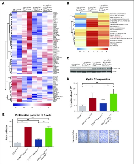
Figure 2.
Gene expression analysis of CD19+B cells from 8-week-old engineered mice. (A) Heat map of the most distinctive sets of differentially expressed genes across indicated genotypes. Values are row-normalized. Cyclin D2 (CCND2) is highlighted in red and validated in panels C and D (see supplemental Table 1). (B) Summary of changes in biological processes and pathways measured by GSEA in CD19+ B cells from mice of indicated genotypes compared with CD19Cre/+ control mice. Color scale represents –log10 false discovery rate (FDR) from GSEA; positive sign reflects correlation to mutant genotype, negative sign reflects correlation to CD19Cre/+ control (see supplemental Figure 3). (C-D) Cyclin D2 expression evaluated by (C) immunoblotting in CD19+ B cells and (D) IHC in the B-cell areas of spleens from mice of the indicated genotypes, calculated by using ImageJ. Bar plots present mean percentage of cells per 5 high-power fields; error bars ± SD. P values were calculated by using Welch’s t test. Representative images are shown below the graphs. Scale bar, 25 µm. (E) Impact of KrasG12D mutation and Ink4a/Arf loss on proliferative potential of CD19+ B cells from mice of the indicated genotype, evaluated by tritiated thymidine ([3H]dThd) incorporation. CD19+ B cells were isolated, stimulated with lipopolysaccharide (20 µg/mL) for 48 hours, and pulsed with [3H]dThd for the final 16 hours of growth. Incorporated [3H]dThd was measured by scintillation counting. Bars represent mean and error bars ± SD of triplicate cultures. P values were calculated by using Welch’s t test. Two independent assays were performed for all experiments for which statistics were calculated. *P ≤ .05, **P ≤ .01, ***P ≤ .001.

