
Fig. 1.
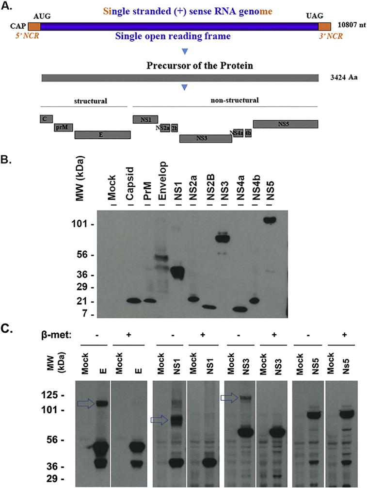
Expression of the ZIKV proteins that are detected by western blot. A. Genomic structure and gene production of ZIKV (MR766 strain, GenBank Sequence Accession: LC002520). AUG: translation start codon; UAG: translation stop codon; NCR: noncoding RNA sequence; nt: nucleotide; Aa: amino acid; B. Western blot assay to examine ZIKV proteins. The ZIKV protein-expressing plasmids were transfected into HEK 293T cells for 24 h. The whole cell lysate samples were applied to run a PAGE and the transferred membrane was blotted with anti-FLAG antibody. The names of the protein were shown on the top and the size marker was shown on the left. C. western blot assay to examine the protein polymerization. HEK293T cells were transfected with the plasmid-expressing ZIKV protein E, NS1, NS3, or NS5 for 24 h. The whole cell lysate samples were collected in a SDS-lysis buffer with or without reducer (beta-mercaptoethanol) and examined for ZIKV protein using anti-FLAG antibody. The experiments have been performed for more than 3 times, one representative is shown.