
Fig. 2.
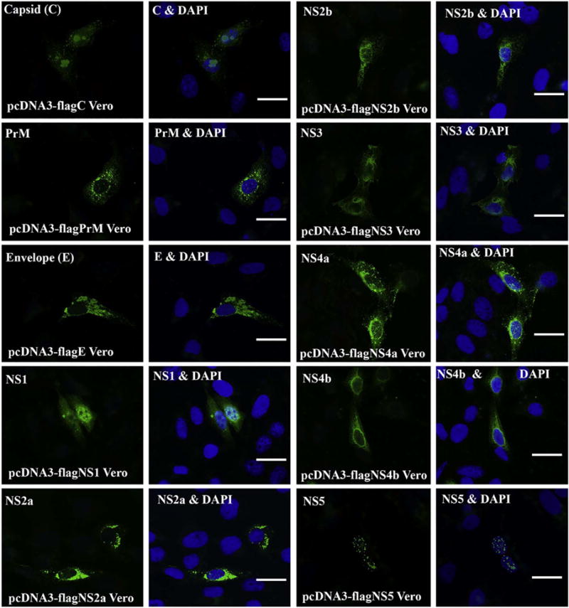
ICC for Visualizing the localization of ZIKV proteins in Vero cells. Vero cells were transfected with the ZIKV protein-expressing plasmid for 24 h. Then, the cells were fixed and permeabilized for ICC. The expressed ZIKV protein was stained with anti-FLAG antibody in green. For each protein, there are two panels, left panel shows the ZIKV protein only in green, and the right panel shows both the nucleus (in blue by DAPI) and the protein. PrM: precursor membrane; C: capsid; E: envelop; NS: nonstructural. Scale bar: 15 μm. (For interpretation of the references to color in this figure legend, the reader is referred to the web version of this article.)