
Fig. 3.
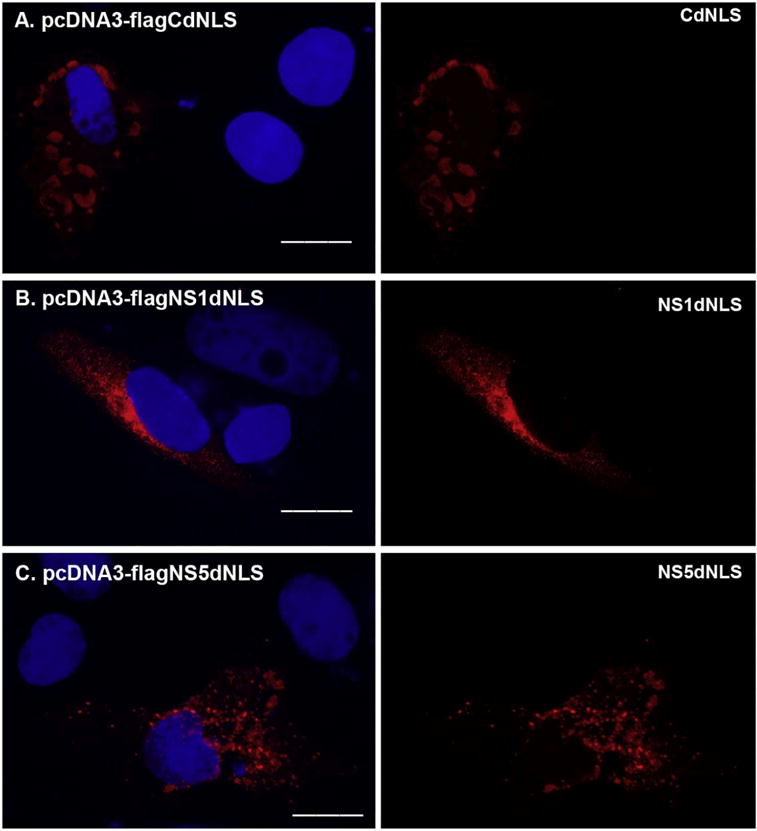
The localization of NLSs mutated ZIKV-encoded proteins. The overlapping PCR method was employed to mutate the putative NLS from protein C, NS1, NS3, or NS5. The primers used for the overlapping PCR were shown in Table 3. The fragments generated by overlapping PCR contain the desired mutation, were gel-purified and cloned into pcDNA3 resulting in pcDNA3-flagCdNLS, pcDNA3-flagNS1dNLS, or pcDNA3-flagNS5dNLS. The plasmid pcDNA3-flagCdNLS (A), pcDNA3-flagNS1dNLS (B), or pcDNA3-flagNS5dNLS (C) was transfected into Vero cells for 24 h. The merged color of the protein (red) and DAPI (blue) was shown in the left panel, and the protein alone (red) was shown in the right panel. ICC was performed to stain the transfected proteins using anti-FLAG antibody in red. DAPI staining was to show the nucleus. Scale bar: 15 μm. (For interpretation of the references to color in this figure legend, the reader is referred to the web version of this article.)