
Fig. 4.
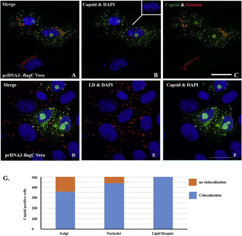
ICC for showing the localization of capsid protein that colocalizes with nucleoli in nucleus and Golgi apparatus or lipid droplet (LD) in cytoplasm. Vero cells were transfected with pcDNA3-flagC. After 24 h, the cells were fixed and ICC was performed to show the relationship between C protein and cellular proteins. A: merged colors of C protein (green), Giantin (Golgi in red) and DAPI (blue); B: merged C (green) and DAPI (blue); C: merged C and (green) and Giantin (red). D: merged C (green), lipid droplet (LD in red) and DAPI (blue); E: merged LD (red) and DAPI (blue); F: merged C protein (green) and DAPI (blue). Scale bar: 10 μm. G. Quantitating the association of C protein with LD, nucleoli, and Golgi apparatus. Five hundred C protein positive cells were counted for the colocalization of C protein with LD, nucleoli, and Golgi apparatus. (For interpretation of the references to color in this figure legend, the reader is referred to the web version of this article.)