
Fig. 6.
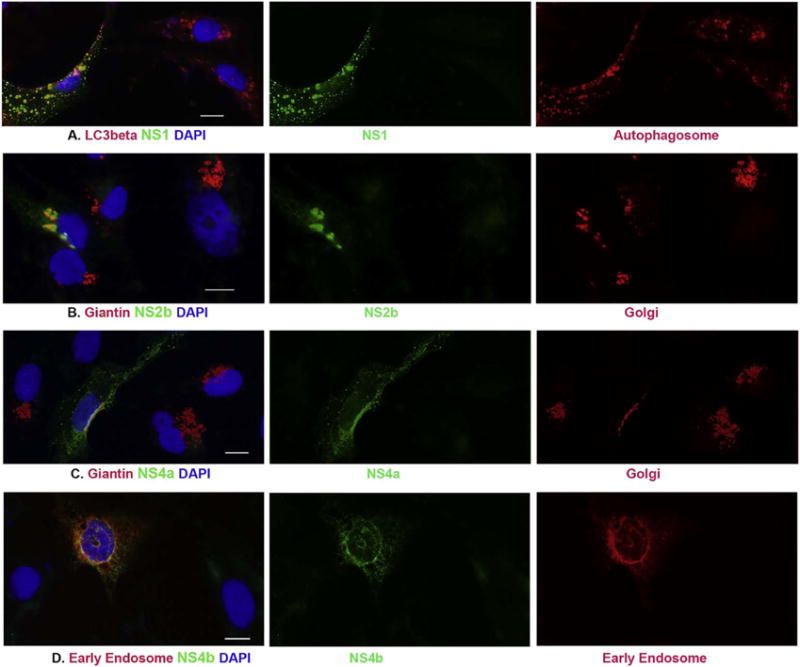
ICC to characterize the localization of NS1, NS2b, NS4a and NS4b localizes in cells after transfection. pcDNA3-flagNS1 (panel A), pcDNA3-flagNS2b (panel B), or pcDNA3-flagNS4a (panel C) was transfected to Vero cells for 24 h. ICC was performed to show the association between ZIKV proteins in green and LC3beta (marker of Autophagosome) or Giantin (marker of Golgi apparatus) in red. D. Vero cells were cotransfected with pcDNA3flagNS4b and pmRFPRab5 for 24 h, anti-FLAG antibody was used show NS4b in green and the early endosome protein Rab5 is in red. The nuclei were shown by DAPI. Scale bar: 10 μm. (For interpretation of the references to color in this figure legend, the reader is referred to the web version of this article.)