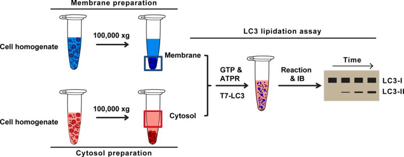
Fig. 1. Cell-free LC3 lipidation assay (modified from (Brier et al., 2016; Ge et al., 2013)).

Membrane preparation: Atg5 KO MEF cells are homogenized and ultracentrifuged at 100,000×g. The membrane pellet is used as the membrane fraction.
Cytosol preparation: WT MEF or HEK293T cells are homogenized and ultracentrifuged at 100,000×g. The supernatant is used as the cytosol fraction.
LC3 lipidation reaction: cytosol, membrane, nucleotides (GTP&ATPR), and LC3-I are incubated for different times. The samples are then analyzed by immunoblotting. The lipidated LC3 appears as a faster migrating band with increased intensity according to time. The LC3-I band shows little change even when more LC3 lipidation occurs in the longer time durations because the overall lipidation efficiency is usually less than 20%.
