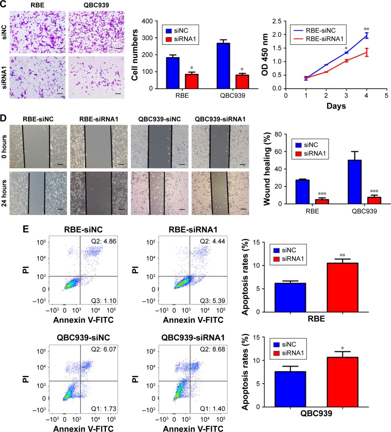
Figure 2.
UHRF2 expression resulted in ICC cell proliferation, invasion, migration, and antiapoptosis.
Notes: (A) UHRF2 expression was interfered by siRNA and confirmed by Western blot and qRT-PCR. GAPDH was used as internal control. (B) Cell counting kit-8 assay was used to assess the ability of cell proliferation. (C) Transwell assay was used to show the invasion of ICC cells with different UHFR2 expression in 48 hours. Scale bar =100 μm. (D) Wound healing assays showed that inhibition of UHRF2 decreased wound healing compared with control cells. Scale bar =100 μm. (E) FCM results indicated that anti-UHRF2 caused acceleration of cell apoptosis. The results are mean ± SD of triplicated independent experiments. *P<;0.05, **P<;0.01, ***P<;0.001.
Abbreviations: FCM, flow cytometry; FITC, fluorescein isothiocyanate; GAPDH, glyceraldehyde-3-phosphate dehydrogenase; ICC, intrahepatic cholangiocarcinoma; qRT-PCR, quantitative real-time polymerase chain reaction; UHRF2, ubiquitin-like with PHD and ring finger domains 2.


