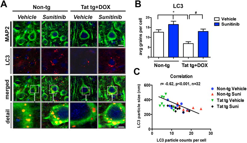
Fig 4. Confocal analysis of the effects of sunitinib on LC3 autophagosomes in neurons of Tat tg mice.
Doxycycline (DOX)-dependent GFAP-Tat tg mice were treated with DOX for 2 weeks to express Tat, and then treated with vehicle or sunitinib for 4 weeks. 8 mice were used per group and were 6.5-7.5 months of age when DOX treatment began. Vibratome sections were double immunolabeled and analyzed by laser scanning confocal microscopy. All representative images are of pyramidal neuronal cells in the frontal parietal cortex. (A) Confocal images of MAP2 (green) and LC3 (red) double labeled neurons showing colocalization of LC3 (orange puncta) in the MAP2-positive cytoplasm of neurons (merged and detail). (B) Computer-aided analysis of LC3-positive puncta size showed sunitinib significantly increased the LC3-positive puncta in non-tg mice compared to vehicle-treated non-tg mice. In vehicle-treated Tat-tg mice LC3-positive puncta size was significantly decreased compared to vehicle-treated non-tg mice, and sunitinib-treatment normalized LC3-positive puncta size in Tat-tg mice. (C) Pearson correlation between puncta size and the number of puncta is inversely related. Vehicle-treated Tat tg mice had a few particles, which were the largest size, while sunitinib-treated Tat tg mice had the most puncta, which were the smallest in size. Statistical analysis performed using ANOVA followed by post hoc analysis using Dunnett's comparison to vehicle-treated non-tg mice (* = p-value < 0.05) or Tukey-Kramer comparison to Tat vehicle-treated tg mice (# = p-value < 0.05). Scale bar = 5 μm, Scale bar in detail panel = 2.5 μm. N = 8 mice per treatment group.

