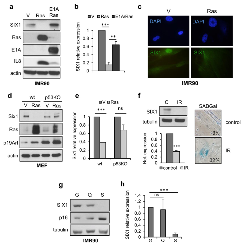
Figure 1. SIX1 regulation during senescence.
(a, b) Western Blot analysis of SIX1 and the indicated controls (a) and QPCR analysis of SIX1 transcript (b) in RasV12-senescent and E1A/Ras (senescence escape) IMR90 human fibroblasts. (c) Immunofluorescence of endogenous SIX1 in Ras-senescent and growing (V) IMR90 cells. (d, e) Western Blot analysis of Six1 and the indicated controls (d) and QPCR analysis of Six1 transcript (e) in RasV12-expressing wild-type MEF (senescent), and p53-KO MEF (senescence escape). (f) Western Blot (top left) and QPCR (bottom left) analysis of SIX1 in senescence induced by gamma irradiation in IMR90 fibroblasts. Representative images and percentage of SABGal positive cells (right). (g, h) Western Blot analysis (g) and QPCR analysis of SIX1 (h) in growing (G), quiescent (Q) and Ras-senescent (Ras) IMR90 fibroblasts.

