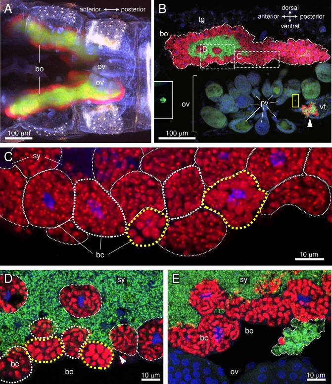
Fig 1. Confocal micrographs of FISH carried out in the abdomen of an adult female D. citri.
Red (Alexa Fluor 594), green (Alexa Fluor 488), and blue (DAPI) signals indicate Carsonella, Profftella, and the host nuclei, respectively. (A) Whole-mount FISH image [maximum intensity projection (MIP)] of the ventral view of the abdomen of an adult female on the day of eclosion showing the bacteriome and immature ovaries. (B) FISH image (MIP) of a sagittal cross-section of the abdomen of an adult female at 5 days post-eclosion. Carsonella can be seen within the uninucleate bacteriocytes on the surface of the bacteriome, while Profftella is encased in syncytial cytoplasm at the center of the bacteriome. Carsonella and Profftella signals can also be seen in an oocyte at the vitellogenic stage (arrowhead), which is located in the ovariole constituting the ovary. Inset on the left is an enlarged image of the area in the yellow rectangle showing a Profftella cell in the hemocoel. (C) Enlarged image (MIP) of the bacteriome in the dotted rectangle of B, from which the Alexa Fluor 488 (Profftella) signals have been removed. Yellow dotted lines surround bacteriocytes containing large spherical Carsonella cells, while thin white dotted lines indicate bacteriocytes containing elongated thin tubular Carsonella cells. Bacteriocytes circled by thick white dotted lines contain Carsonella cells that appear to be in the process of transformation. (D) Enlarged image (optical section) of the area shown in B. Dotted lines indicate the same structures as described in C. The arrowhead indicates the syncytium harboring Profftella, which reaches the surface of the bacteriome. (E) FISH image (MIP) of another sagittal cross-section of the abdomen of the same individual shown in A–D. A mass of spherical Carsonella and Profftella cells exiting from the bacteriome is indicated within the dotted line. Abbreviations: bc, bacteriocyte; bo, bacteriome; ov, ovary; pv, previtellogenic oocyte; sy, syncytium; tg, tergite; vt, vitellogenic oocyte.

