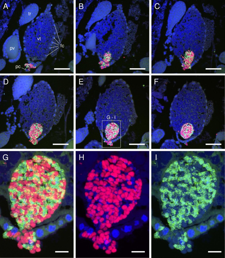
Fig 2. Confocal micrographs (MIP) showing transovarial transmission of Carsonella and Profftella.
(A–F) FISH images showing an oocyte accepting symbionts in serial sections (5 μm thick) of an adult female D. citri at 5 days post-eclosion. Red (Alexa Fluor 594), green (Alexa Fluor 488), and blue (DAPI) signals indicate Carsonella, Profftella, and the host nuclei, respectively, unless otherwise stated. A mass of Carsonella and Profftella cells can be seen entering the posterior of a vitellogenic oocyte through follicle cells and the pedicel of the ovariole. (G) Enlarged image of the dotted rectangle shown in E. (H) Duplicate of image shown in G following the removal of Alexa Fluor 488 (Profftella) signals. Spherical or short rod-shaped Carsonella cells can be seen entering the posterior of the oocyte through follicle cells. Note that the shape of the Carsonella cells does not change after entering the oocyte. Some of the weak DAPI signals in this micrograph correspond to Profftella (see also I). (I) Duplicate of the image shown in G following the removal of Alexa Fluor 594 (Carsonella) signals. Spherical Profftella cells can be seen entering the posterior of the oocyte through follicle cells. Note that Profftella cells in the oocyte have already transformed from spherical to tubular form. Some of the weak DAPI signals in this micrograph correspond to Carsonella. Abbreviations: fc, follicle cell; pc, pedicel; pv, previtellogenic oocyte; tr, trophocyte; vt, vitellogenic oocyte. Scale bars: 50 μm in A–F, 10 μm in G–I.

