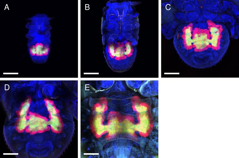
Fig 4. Confocal micrographs (MIP) of whole-mount FISH assays showing the bacteriomes of the D. citri nymphs.
Red (Alexa Fluor 594), green (Alexa Fluor 488), and blue (DAPI) signals indicate Carsonella, Profftella, and the host nuclei, respectively. Bar: 100 μm. (A) 1st instar. (B) 2nd instar. (C) 3rd instar. (D) 4th instar. (E) 5th instar. The bacteriome continuously increased in size and volume during nymphal development in proportion to the increase in body size. The pair of wing-like protrusions that emerged at the late embryonic stage continued to grow throughout the nymphal stages.

