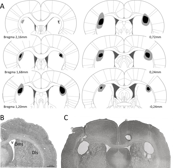
Fig 1.
A. Reconstruction of the dls lesions displayed on standard coronal sections from the atlas of Paxinos and Watson [35]. The largest lesion is shown in pale shading and the smallest in dark shading. B. Photomicrograph showing a no lesioned coronal brain section and (C) a coronal section after excitotoxic lesion of dls. Dls: dorso-lateral striatum; Dms: dorso-medial striatum; v: lateral ventricle.

