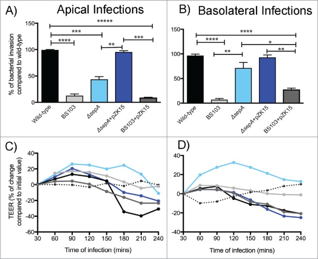
Figure 1.

SepA facilitates bacterial invasion by opening the epithelial barrier. Bacterial invasion was assessed by infecting polarized epithelial cell monolayers either at the apical (A) or basolateral (B) poles with the following S. flexneri strains: wild-type (black), an avirulent, non-invasive BS103 (light gray), ΔsepA (light blue), ΔsepA+pZK15 (dark blue), and BS103+pZK15 (dark gray). Data are expressed as colony-forming units (CFU) per monolayer ( ± SEM) compared with wild-type. Regulation of TEER after stimulation with S. flexneri strains was also evaluated after infecting the apical (C) or basolateral (D) poles with the same strains used above. TEER values are expressed as percentage of change compared with initial values. The data are expressed as means ± SEM of triplicate samples for all conditions tested.
