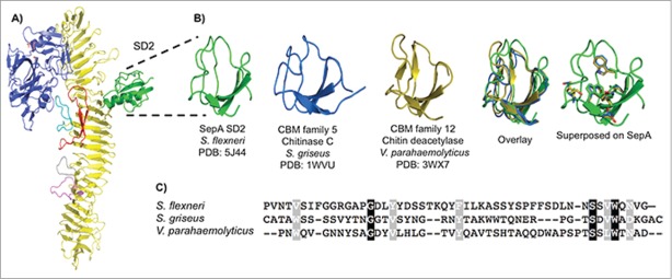
Figure 6.
SepA SD2. (A) SD2 highlighted in a ribbon representation of SepA. (B) Side-by-side comparison of minimal SepA SD2 (residues 491–541, green) with chitinase C, a carbohydrate-binding module 5 family member from S. griseus (residues 32–79, blue), and chitin deacetylase, a carbohydrate-binding module family 12 member from V. parahaemolyticus (residues 333–379, orange). Overlay of the 3 truncated proteins and superimposition of conserved residues from the protein alignment onto SepA. (C) CLUSTALW alignment of the 3 aforementioned amino acid sequences. Conserved residues are shown in black and semi-conserved residues in gray.

