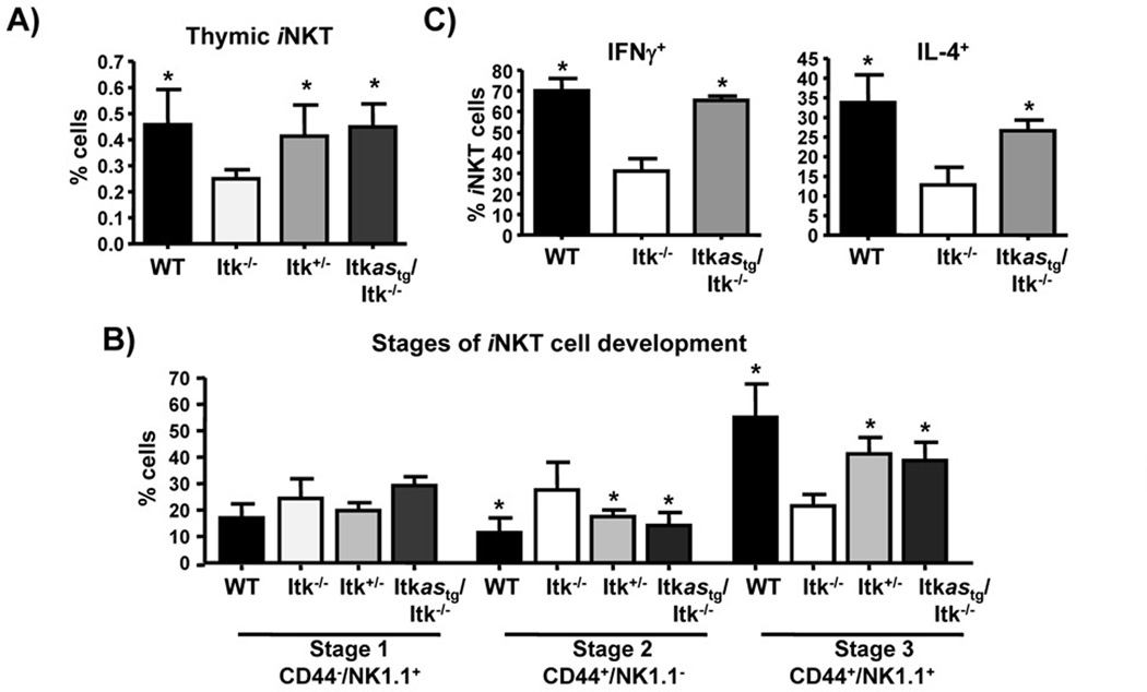
Figure 4. Itkas rescues iNKT cell development.
A) Thymocytes from the indicated mice were analyzed for percent of iNKT cells by gating on CD1d tetramer+TCRβ+ cells. (B) Thymic iNKT-cells were analyzed for expression of CD44 and NK1.1 and divided into Stage 1, Stage 2, and Stage 3 according to CD44 and NK1.1 expression. Data are shown as mean ± SEM of n= 5 mice/group, representative of at-least two independent experiments. *p < 0.05 by unpaired Student t-test. (C) WT, Itk−/− and Itkastg/Itk−/− mice were injected with 2 µg of α-GalCer. Two hours later, spleens were isolated and analyzed for production of IFN-γ and IL-4 by iNKT cells using flow cytometry.

