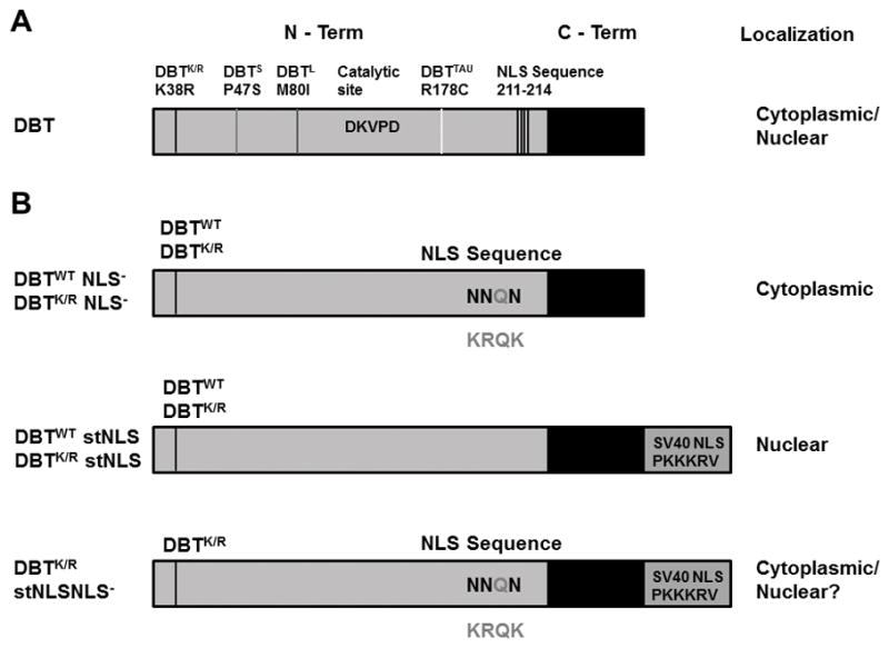
Figure 1.

Domain structure of DBT and sites of mutations.
A) Location of previously characterized period-altering mutations of dbt, the catalytic site and the NLS sequence. The NLS mutation is away from the catalytic region of DBT. B) DBT mutant proteins analyzed in this study. The residues outside the proteins indicate the amino acids that are part of the putative NLS sequence. The residues in the NLS mutated to Asparagine are the darker ones, as shown inside the protein. The addition of strong SV40 NLS sequence is shown below the NLS− mutant. Predicted subcellular localizations are shown to the right; the DBTK/R stNLS NLS− mutant was predicted to be nuclear because of the addition of the stNLS but in fact is less nuclear than the DBTK/R stNLS protein (hence the “?”),.
