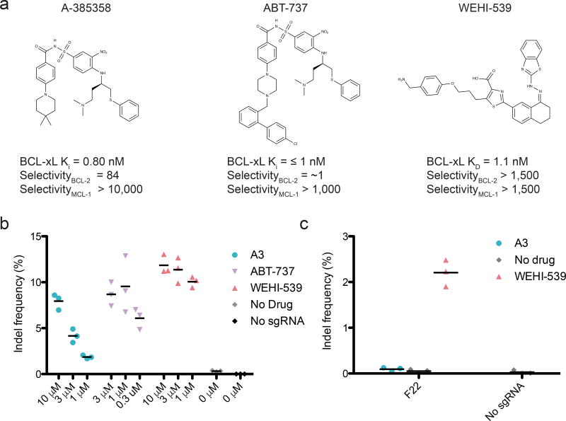
Figure 5. ciCas9 can be activated by a variety of BCL-xL disruptors.
(a) Structures of the three BCL-xL disruptors used in this study are shown with their reported Kd or Ki for BCL-xL, and their selectivity for BCL-xL over BCL-2 or MCL-137,40–42. (b) Editing at the AAVS1 locus 24 hours after ciCas9 activation with different concentrations of the three disruptors is shown. The A3 data are also shown in Fig. 1c. Black bars depict means (n = 3 cell culture replicates). (c) Editing is shown at the AAVS1 locus 24 hours after activation of the ciCas9(F22) variant with 10 µM of each disruptor. Black bars depict means (n = 3 cell culture replicates).

