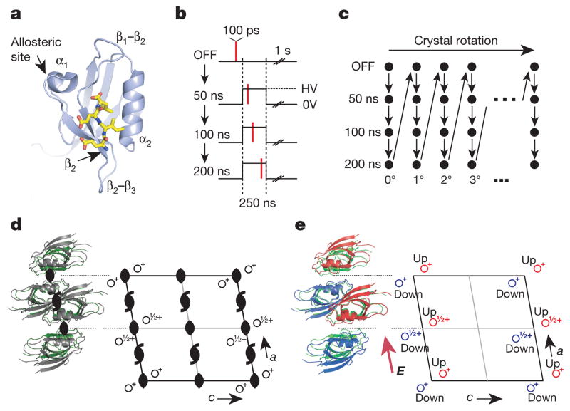
Figure 2. An EF-X experiment in the LNX2PDZ2 domain.
a, LNX2PDZ2 binds target ligands (in yellow) in a groove between the β2 and α2 segments. The binding site is coupled to allosteric sites on the β2–β3 segment and the α1–β4 segment (through the β1–β2 loop and α1 helix). b, Data collection involves four sequential X-ray exposures for each crystal orientation: no voltage (OFF), and three time delays (50, 100, 200 ns) after onset of the voltage pulse. One second is allowed between pulses for crystal cooling. HV, high voltage. c, The protocol in b is repeated for a series of crystal rotations to collect a full diffraction data set. d, LNX2PDZ2 crystallizes in the C2 space group, which includes two kinds of rotational symmetry (black symbols); this results in four molecules per unit cell and one molecule per asymmetric unit. e, With the electric field E (applied along the a dimension), all rotational symmetry is broken. This results in a new unit cell with two molecules per asymmetric unit (red and blue)—one experiencing + E, and one experiencing − E.

