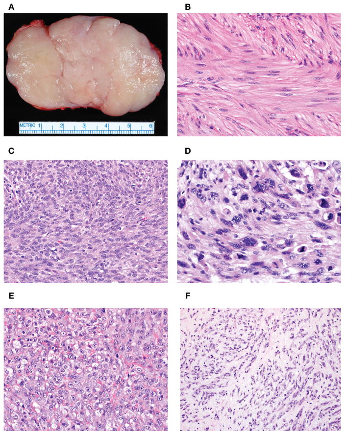
Figure 1.
Vulvovaginal SMTs are often conservatively excised or enucleated in an absence of clinically concerning features for malignancy given their proximity to sensitive anatomic structures (A). This surgical procedure often results in problematic microscopic assessment of tumor interface (infiltrative or circumscribed). SMTs with mild cytologic atypia exhibited minimal variation in nuclear size and shape, stippled and evenly dispersed chromatin and small to inconspicuous nucleoli (B). Tumors with moderate cytologic atypia had larger nuclei with irregular nuclear membrane contours, uneven chromatin and more prominent nucleoli (C). Tumors with severe atypia demonstrated significant nuclear enlargement and pleomorphism with coarse chromatin and large nucleoli (D). Epithelioid morphology showed polygonal to rounded cells with a nested or sheet-like growth (E). Myxoid morphology featured prominent quantities of myxoid acid-mucin stroma that often resulted in dyscohesion or separation of individual cells (F).

