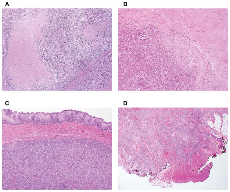
Figure 2.
Tumor cell necrosis exhibited an abrupt transition between viable to non-viable tumor without evidence of intervening tissue (A). In contrast, ischemic necrosis showed a zone of granulation tissue and/or hyalinization between viable and non-viable tumor (B). Tumor interface was classified as circumscribed when it was well-demarcated relative to surrounding non-neoplastic tissue or had an expansile growth (C) and as infiltrative when permeative growth or destructive invasion of surrounding tissue was present (D).

